Low-grade bladder tumors are almost always confined to the urothelial surface. They are arguably the most common urothelial tumor in the urinary tract. The current grading system uses low and high grade instead of the now historical 1–3 grading system. Low-grade tumors include not only those that were previously called grade 1 but also some of the grade 2 tumors. If the pathologist believes there is “significant” atypia they will classify the bladder cancer as high grade. Thus high grade incorporates not only all the prior grade 3 but also many of the prior grade 2 tumors. In an analysis of a 10 year span of pathology reports from The University of Miami we demonstrated a significant decline in reports of low grade bladder cancer with a corresponding increase in those called high grade. In other words there has been a grade migration. This is important as it may lead to overtreatment for some patients.
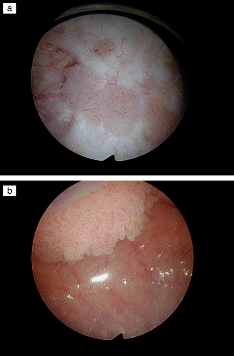
Fig.5
Papillary bladder tumors.
The current EAU and SUO/AUA guidelines state that patients with low risk bladder tumors have a single initial papillary low grade tumor less than 3 cm. Any case of primary multifocal low grade Ta bladder cancer or any patient with “recurrent” low or high grade Ta tumor regardless of size are in the intermediate risk category. If we believe the risk category should classify patients according their risk of progression then all patients with low-grade tumors should be low risk since they rarely develop an invasive bladder cancer. They may be at intermediate risk for a “recurrence” if they have multifocal or large tumors but they are at very low risk of progression, which, in my view, is the most important prognostic factor in determining treatment decisions.
Fig.6
Papillary tumor in the prostatic urethra.
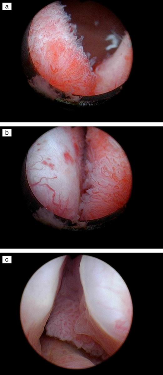
Urothelial cancer in the prostatic urethra is relatively uncommon. Grade and depth of invasion are critical as it relates to the extent of resection and consideration of subsequent treatment. The current case involves a man with low-grade non invasive appearing tumor in the prostatic urethra in addition to tumors in the bladder. We invite our readers to review and comment on the case and management by using the online comment section below the case: https://www.bladdercancerjournal.com/challenging-cases.
Case
This is a 70 year old man with a heavy cigarette smoking history and despite stopping at age 50 he has moderately severe chronic obstructive pulmonary disease (COPD) as well as a history of recurrent low and high-grade non invasive (Ta) bladder cancer. The most recent outpatient flexible cystoscopy identified several papillary Ta appearing tumors in the bladder and similar tumors on the surface of the prostatic urethra.
He has minimal lower urinary tract symptoms (LUTS). He gets up once at night to void and does not have daytime frequency. His post void residual urine is 32 ml.
He has undergone a formal transurethral resection of bladder tumors (TUR BT) approximately every two years over the past decade. In addition to formal TUR BTs I have cauterized small papillary low-grade Ta appearing tumors in the office on several occasions. During the most recent office cystoscopy I observed several small and medium size bladder tumors and also papillary low grade appearing tumor in the prostatic urethra.
In the preoperative holding area prior to surgery I reviewed my checklist and as an important part of that I discussed the case with the attending anesthesiologist. He told me that the patient was having an acute exacerbation of bronchitis and he did not feel a general anesthetic was appropriate however the patient agreed to a spinal anesthetic. I consented the patient for a TUR BT as well as a transurethral resection of the prostate (TURP).
I realize there are several alternatives for eradicating the apparent “superficial” tumor in the prostatic urethra (PU). This includes a formal TURP, laser or roller ball cauterization, or cold cup biopsy followed by cautery.
I began the procedure by inserting a continuous flow resectoscope using a visual dilator. I used a 70 lens to review the entire bladder and confirmed the presence of several papillary bladder tumors. They ranged from 1–3 cm and were located on the posterior and anterior walls. The papillary tumor in the PU was present at the bladder neck/median lobe of the prostate, along sections of both lateral walls and near the verumontanem.
I removed the bladder tumors by a standard TUR BT using the monopolar resectoscope. Given the current appearance of the tumors and his history of only having Ta tumors I made no attempt to resect beyond the lamina propria. I proceeded to perform a limited TURP. I felt confident that the tumors were low grade and almost certainly noninvasive. Following the procedure a catheter was placed and the patient was discharged home and instructed to remove the catheter in 2 days.
Would you have proceeded in a different fashion?
Would you give him postoperative intravesical chemotherapy?